Articular surfaces of the femoral condyles
In order for the knee joint to glide smoothly and painlessly, the cartilage covering on the femoral condyles and the tibial head must be intact. Here, cartilage damage is mainly found on the inner side of the joint. Depending on the extent of the damage, different therapy methods can be used:
MICROFRACTURE SURGERY
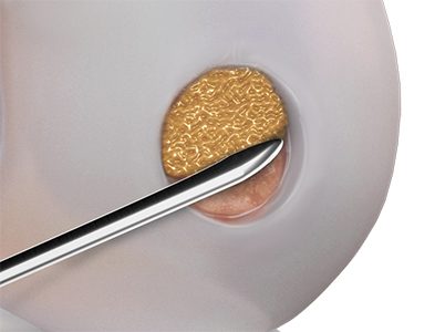
Fine holes are drilled into the bone with a type of awl. The concept is the same as the Pridie technique: the sclerosed bone is pierced, stimulating the blood vessels at the area. The ensuing influx of mesenchymal stem cells stimulates the formation of new cartilage. Research shows a very encouraging success rate for this technique - as high as 80%.
The advantage over Pridie drilling is that microfractures are created using a special awl, whereas the high speed of the drill used in the Pridie procedure can generate considerable heat, burning the bone and blood vessels. With microfracturing, no heat damage is incurred.
Dr. Gäbler achieves excellent outcomes using the microfracture technique; often, cartilage damage is no longer noticeable after just a few months. For this reason, Dr. Gäbler prefers this method of treating relatively minor cartilage damage, rather than a cartilage cell transplant (which is much more complex).
Find out more about knee injuries in sports in this Kurier interview with Dr. Gäbler (available in German only).
This arthroscopic image shows the holes that remain following microfracturing.
MOSAICPLASTY
Smaller cartilage defects in the weight-bearing areas of the knee can be repaired using a mosaic-like grafting technique that harvests cartilage from parts of the knee where it is less needed (non-weight-bearing areas). These cartilage plugs are then inserted into the defective areas, where they are able to heal into place.
However, this method can leave behind tunnels (lifting defects), which often cause pain—the method is therefore only used in exceptional cases (see below).
What is mosaicplasty best suited for?
Mosaicplasty is a very good method for treating Ahlbäck’s disease (aseptic bone necrosis) and OCD (osteochondrosis dissecans) of the knee and ankle.
CARTILAGE TRANSPLANT (MACT)
Larger defects can be treated using cartilage transplants. To do this, an operation is first carried out to harvest cartilage, which is then cultivated in a cell culture lab under sterile conditions. About six weeks after the initial operation, the newly grown cartilage can be used as a transplant in a second operation. Cartilage transplants are indicated in only a very limited number of cases, as there must be no meniscus damage and cartilage must be intact on at least one of the two joint surfaces.
AutoCart Cartilage (MINCED CARTILAGE)
A simpler method that requires only a single operation is a cartilage graft using the AutoCart procedure (Arthrex). This method is useful for grafting more significant cartilage damage in what is usually a minimally invasive procedure.

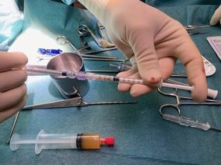
With the AutoCart method, cartilage is shaved from the defective edges and immediately reduced is size (minced). As the edges of the cartilage must be straightened anyway, no further cartilage damage is caused (as is the case, for example, with mosaicplasty), only in rare cases is it necessary to remove cartilage from other non-weight-bearing areas. The cartilage is then mixed with endogenous PRP (ACP) and thrombin, creating a paste that is introduced into the defective area, and coated with thrombin and ACP to create a seal. The ACP contains high doses of growth factors that stimulate cartilage healing. Thrombin is a crucial macromolecule for blood clotting and is taken from the patient’s blood during the operation.
The main advantage of this method is that only substances from the patient’s own body are used, meaning that there can therefore be no rejection and only a single operation is needed.

In most cases, the patient is discharged one or two days after the operation at the most. An orthosis (splint) must be worn for six weeks and mobilization with crutches is likewise required for six weeks to avoid overstressing. Physiotherapy is essential to the healing success.
This procedure is a recognized treatment method with very good therapeutic results. It was decisively improved by the Arthrex company through their development of new instruments and the use of ACP (PRP) and thrombin.
CARTILAGE DEBRIDEMENT
Dr. Gäbler often sees patients with a history of arthroscopy that included cartilage smoothing, or cartilage debridement. In almost all cases, patients felt worse after cartilage smoothing than before.
Scientific studies now clearly show that cartilage debridement (especially in older patients) not only fails to lead to improvement, but often even causes symptoms to worsen.
As Dr. Gäbler explains, the problem is that the cartilage smoothing process also grinds away good cartilage; this is because the differences between the types of cartilage cannot be distinguished during arthroscopy.
Some Austrian and German medical associations still recommend cartilage smoothing, but Dr. Gäbler considers the method to be a medical failure and advises his patients against the procedure.
A clear exception is in cases requiring the removal of cartilage scales, which could otherwise loosen and detach, causing pieces to freely float around the joint.
AUTOLOGOUS BLOOD TRANSFUSION (ACP)
ACP (autologous conditioned plasma) is an autologous blood product. Blood is taken from a vein in the patient’s arm and centrifuged using a specialized technique. This activates the platelets, which in turn release proliferative substances (such as platelet-derived growth factor (PDGF) and morphogenic proteins (transforming growth factor, TGF), which are key to stimulating muscle, tendon, cartilage, and bone healing.
You can find more information about ACP therapy at the SPORTambulatorium Wien here.
CINGAL GEL INJECTIONS - CROSS-LINKED HYALURONIC ACID
Cingal gel contains cross-linked, stable hyaluronic acid, which has been extensively studied. Hyaluronan, or sodium hyaluronate, is a sugar-like molecule also called a polysaccharide. The molecular structure of hyaluronan, an important component of synovial fluid, is a long chain made up of many identical links. Hyaluronic acid is created by cells in the synovial membrane and released into the joint cavity, where it helps lubricate the surface of the cartilage. The length of the hyaluronan chains affects the lubricating properties (viscoelasticity) of the corresponding medications.
SIDE EFFECTS OF CINGAL GEL
Localized side effects such as pain, heat, redness, and swelling may accompany treatment of the joint. This is a very common side effect of cross-linked hyaluronic acid and, although such reactions are unpleasant, the knee generally calms down within just a few days. In rare cases, hypersensitivity has been observed.
ACTIVE MECHANISM OF HYALURONIC ACID
It is not known precisely how hyaluronic acid works, but research has shown that arthritic joints have significantly lower concentrations of hyaluronic acid than healthy joints. Injecting hyaluronic acid into the affected knee joint increases its lubrication. Hyaluron also has an anti-inflammatory effect.
Broad studies have shown that cross-linked hyaluronic acid infiltrations are more effective than injections of simple hyaluronic acid. Additionally, simple hyaluronic acid has the disadvantage of requiring 3-5 injections into the knee, usually one a week, while a single shot of cross-linked hyaluronic acid is often effective for several months.
The gel implantation of cross-linked hyaluronic acid improves natural joint function, extending the joint’s longevity by restoring the mechanical function of the natural synovial fluid (viscoelasticity). In addition, Cingal reduces certain enzymes that cause cartilage deterioration, thus slowing the process. The high stability of Cingal gel means that a single treatment is often effective for 6-9 months.
Cost per infiltration: € 350
More questions?
Our experts are happy to help you
Just give us a call!


