
Seitenbandverletzungen können isoliert oder als Kombinationsverletzung, vor allem in Kombination mit Verletzungen des Meniscus and Kreuzbänder auftreten. Isolierte Seitenbandverletzungen und knöcherne Ausrisse finden sich vor allem bei Kindern. Diese Verletzungen können meist problemlos mit einem Gips oder einer Schiene ausbehandelt werden.
In adulthood, it is essential to rule out any secondary injuries.
Isolated injuries to the inner collateral ligament can be immobilized with a splint and, depending on the severity, exercise therapy should begin as quickly as possible. Surgical treatment of isolated collateral ligament injuries is required only in rare cases.
Collateral Ligaments: Function
The knee joint has an inside (medial) and an outside (lateral) collateral ligament. The medial collateral ligament runs from the inside of the thigh to the shin, in close proximity to the base of the medial meniscus. The lateral collateral ligament runs from the outside of the thigh to the head of the fibula.
The collateral ligaments provide passive stability to the knee, preventing valgus (knock knees) and varus (bowleg).
Collateral Ligament Injuries
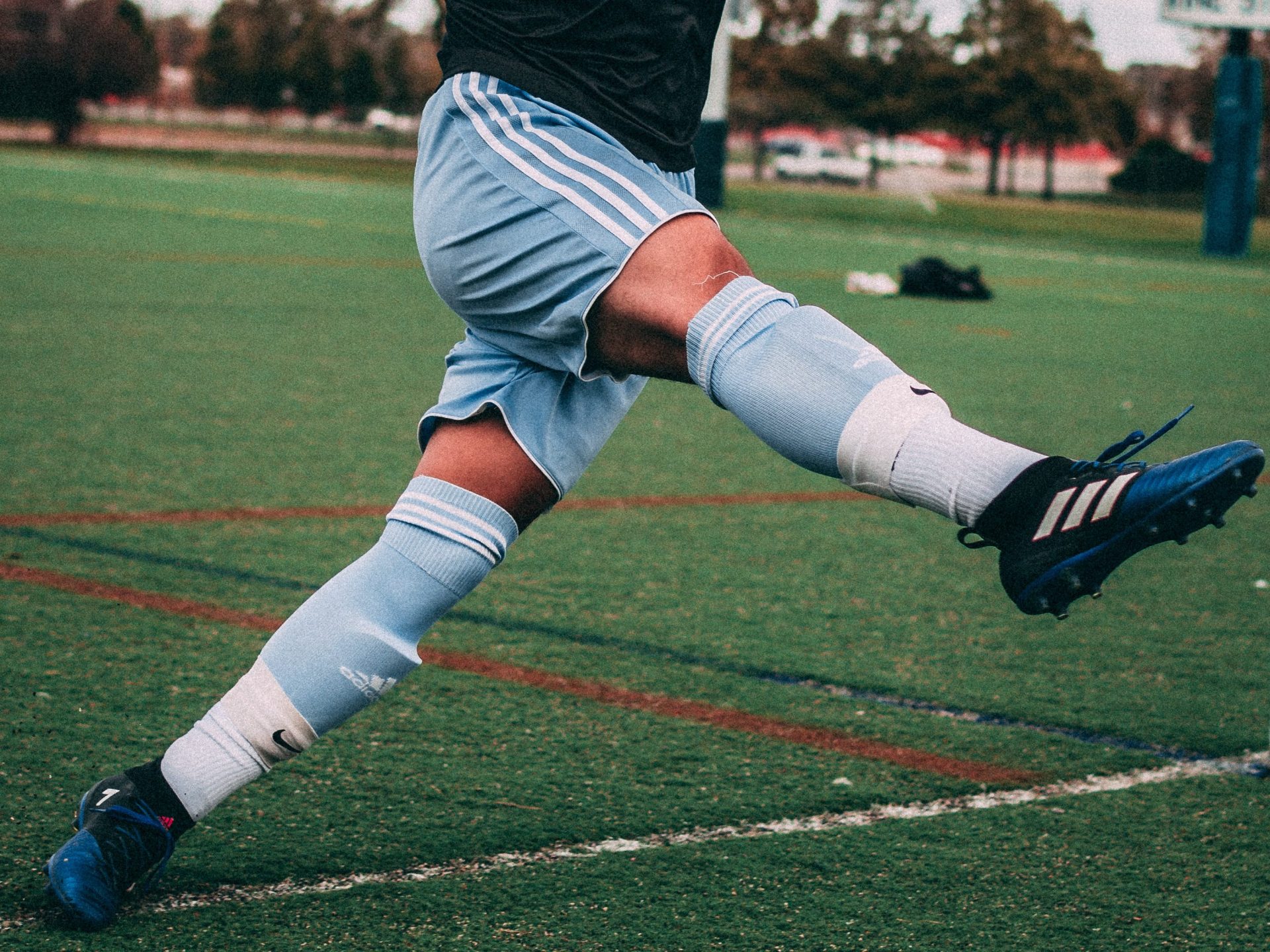
The most common cause of a collateral ligament injury is an accident while playing sports. Depending on the intensity of a lateral impact, it can cause a strain, partial rupture, full rupture, or an avulsion fracture. Medial ligament injuries are more common than lateral ones.
Symptoms
The primary symptom is pain around the injured ligament, in most cases accompanied by swelling and bruising.
If the collateral ligament is severely or fully ruptured, the patient often also experiences a sensation of instability, especially when under stress, accompanied by pain and the formation of a hematoma around the rupture.
Common associated injuries include meniscal tears, and in some cases cruciate ligament injuries, which are accompanied by the corresponding symptoms.
Due to what is often severe pain and the feeling of instability, we recommend going to the nearest hospital at the time of the accident if at all possible. There, a preliminary diagnosis and immobilization the injury with a splint will be carried out (if required).
Examination
The first step is to record the exact time of the accident and how the injury occurred (anamnesis). This is documented with precision, particularly in the case of leisure, sports, and work accidents, in order to make sure there are no difficulties with insurance coverage. This is followed by a clinical examination of the injured knee joint. The contusion, pain level, range of motion and - most importantly - ligament stability are examined with care. A stress test is carried out to determine the range of the affected ligament, with gapping divided into three degrees:
- Grade 1: less than 5mm – slight instability
- Grade 2: 5 to 10mm – medium instability
- Grade 3: more than 10mm – severe instability
Im Krankenhaus wird meistens ein Röntgen durchgeführt, um eine knöcherne Verletzung des Kniegelenkes auszuschließen. Dringend angeraten ist bei Seitenbandverletzungen aber in weiterer Folge auch eine MRI Untersuchung. Hiermit kann bildgebend nachgewiesen werden, wie groß das Ausmaß der Verletzung ist. Mittels MRI können auch Begleitverletzungen wie Meniskusriss, Cruciate Ligament Rupture (ACL Tear), Knorpeldefekte und Bone Bruises diagnostiziert werden.
Treating a Collateral Ligament Injury
The first steps in treating the injury are rest, icing, and elevation. Massive swelling can be prevented by wrapping with a compression bandage.
If there is a feeling of instability, the patient should walk with the support of crutches and/or a splint even before visiting a doctor, especially if such aids are available. However, please remember the absolute necessity of preventing thromboembolism when resting the injured leg!
A medical diagnosis will indicate if conservative or surgical is indicated based upon the degree of injury and presence of secondary injuries:
Conservative treatment:
- Cool, rest
- Pain medication
- Compression bandage
- Knee brace for 4–8 weeks (for mild to moderate instability)
- Mobilization with crutches
- Physical therapy as soon as possible
Surgical treatment options (arthroscopic or minimally invasive surgery):
Collateral ligament suture
After a knee arthroscopy to directly assess the extent of the injury and treat any secondary injuries, the torn sections of the collateral ligament can be surgically sutured.
Refixation of an avulsion fracture
After a knee arthroscopy to directly assess the extent of the injury and treat any secondary injuries, an anatomical repositioning and fixation of the bone fragment can be carried out. Depending on the size of the fragment, this is done either with suture anchors or osteosynthesis screws.
Ligament augmentation with InternalBrace™ by Arthrex
After a knee arthroscopy to directly assess the extent of the injury and treat any secondary injuries, the torn parts of the collateral ligament are surgically sutured. This is then reinforced using an internal brace. The InternalBrace™ augmentation of the medial collateral ligament uses a 2-mm-wide FiberTape suture between two knotless SwiveLock anchors. The brace protects and reinforces the primary collateral ligament suture and is indicated when the degree of instability is high or when mobilization without a brace is required.
- Operation duration: approx. 45 minutes
- Scar length: approx. 3–4cm, arthroscopic incisions approx. 6mm
- Suture type: cosmetic seam, thread removal not necessary
- Anesthesia: general or spinal anesthesia possible
- Hospital stay: 1–2 nights
- Physical therapy: begins immediately post operation
- Follow-up at the SPORTambulatorium: 2 weeks after surgery
More questions?
Our experts are happy to help you
Just give us a call!