Heel Spurs
A heel spur is a thorn-like growth on the bone at the bottom of the calcaneus, or heel bone.
Typical symptoms are stabbing pain on the bottom or side of the heel, especially in the morning and when first getting going.
The trigger is inflammation of the plantar aponeurosis (plantar fasciitis). This is caused by prolonged overstressing through athletic activity, as well as by poor footwear, incorrect weight bearing, misalignment, and obesity.
Conservative therapy includes wearing insoles, anti-inflammatory medication, taking a break from sports, and shockwave therapy, all treatments that will soon lead to improvement. Surgery should only be considered if other therapy methods do not achieve the desired results.
More questions?
Our experts are happy to help you
Just give us a call!
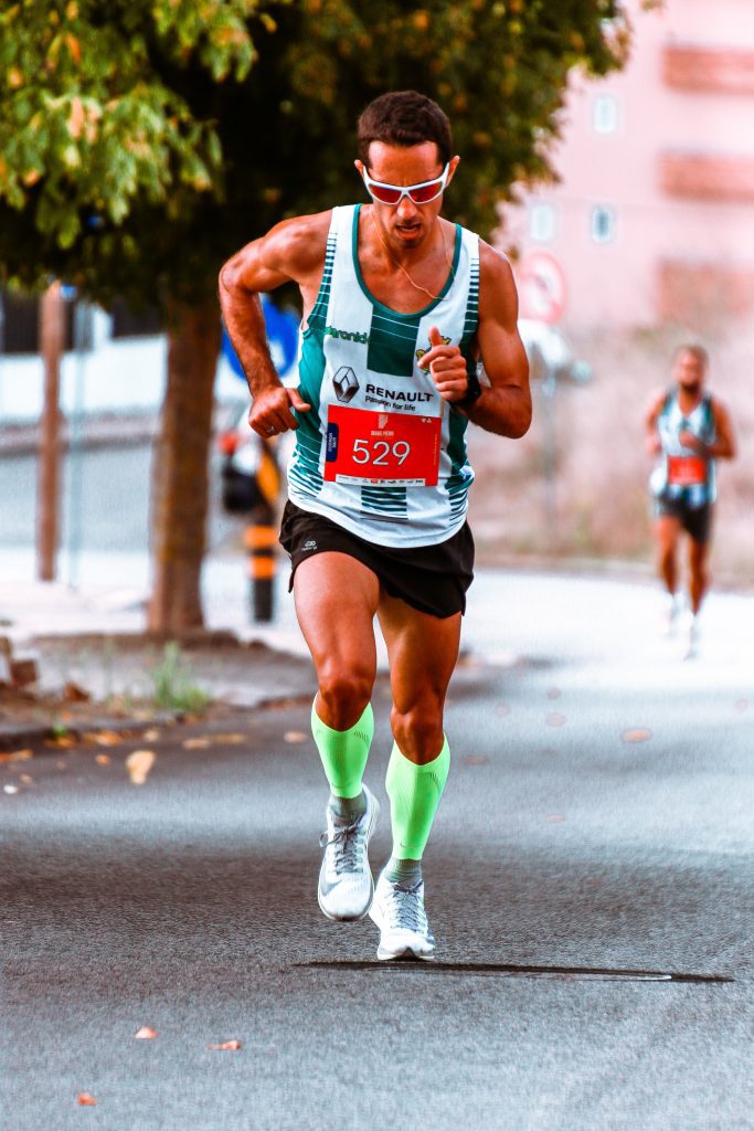
Dr. Gäbler is frequently approached by running enthusiasts who complain about discomfort in the heel area. Some say that they occasionally wake up to pain so severe that it feels like they have stepped on a nail. But then, after a few limping steps, it usually becomes possible to walk normally again.
The trigger for these symptoms is often a heel spur, inflammation of the plantar aponeurosis (plantar fasciitis). This is caused by prolonged overstressing of the foot and tendons resulting from excessive pressure and tension on the aponeurosis at the sole of the foot underneath the heel bone. Repeated micro-tears of the tendon and chronic inflammation of the surrounding tissue occur at these overstressed areas, causing calcium deposits to develop. (As with a bone fracture, the body deposits calcium to heal the tears in the tendon and provide stability.) And just like that: a bone spur. Similar processes are also found in tennis elbow, another frequently occurring periosteum inflammation.
Clear ossification does not always have to be present in order to experience the symptoms of a heel spur, as the pain is caused by a chronic inflammation that spreads to the bursa of the calcaneus.
A calcaneal spur visible on an X-ray occurs in around 10% of the population, however the majority of people have no symptoms. Pain in the region of the calcaneal spur, however, can occur as a result of overexertion (age, obesity, running).

Anatomy:
The bones of the foot have an arched shape. The plantar aponeurosis stretches the arch of the foot like a cord, preventing it from becoming flatter. The plantar aponeurosis, also known as plantar fascia, is fibrous and originates in the mid section of the calcaneus. From there, it divides into ligaments attached to the toes. A failure to function can very rapidly lead to the arch of the foot collapsing, causing a flat foot, midfoot pain, and later even to arthritic joints.
Causes:
The trigger for these symptoms of heel spurs is inflammation of the plantar aponeurosis (plantar fasciitis). This is caused by prolonged overstressing of the foot and tendons resulting from excessive pressure and tension on the aponeurosis at the sole of the foot underneath the heel bone. Repeated micro-tears of the tendon and chronic inflammation of the surrounding tissue occur at these overstressed areas, causing calcium deposits to develop. (As with a bone fracture, the body deposits calcium to heal the tears in the tendon and provide stability.) And just like that: a bone spur. Similar processes are also found in tennis elbow, another frequently occurring periosteum inflammation.
Heel pain can occur in obese patients as well as very active people and athletes, supporting this explanation. Patients with a fallen arch, splayed feet, or with high arches also have a tendency to experience heel pain.
Similarly, a spur can also form in the context of other inflammation (e.g. in rheumatic diseases). A fallen arch can cause a heel spur to form by changing the position of the calcaneus and causing pressure on the tendon's anchor point.
The most common causes, however, are poor footwear and excessive or incorrect loading of the footcaused, for example, by the malpositioning of flat feet. Long periods of standing and running on hard surfaces, obesity, insufficient warm-up periods during sports, and shrinking of the fat pad at the heel due to age are all factors that contribute to the development of a heel spur.
Symptoms:
- Severe stabbing pain beneath or on the side of the heel when standing or walking.
- Pain that subsides when the foot is not in use and is more severe upon standing up, particularly in the mornings. When the foot is relaxed (e.g. at night), the bursa swells significantly. The first steps are then especially painful.
- Symptoms are considerably worse when running long distances. Running on hard surfaces is also painful.
Diagnosis:
A heel spur can grow up to 10 mm in size and be examined by palpation, as pressure at the center of the calcaneus triggers pain. An X-ray is often made in order to rule out any other diseases such as rheumatism or tumor—or even a stress fracture. of the heel bone.
If a calcaneal spur is not visible on the X-ray, an MRI can reveal signs of chronic inflammation (plantar fasciitis).
Who is particularly at risk?
- Long distance runners
- Middle-aged weekend runners, as the shock-absorbing pad at the heel shrinks over the years
- Obese patients
- Persons with an uncorrected malpositioning of the foot.
Treatment:
In terms of treatment, surgery is generally the very last resort. Non-surgical treatment options often quickly lead to success.
- Inserts relieve the painful point on the heel by preventing direct contact of the heel with the floor when walking. Preferably in combination with physiotherapy (stretching exercises - these can also be carried out at home: e.g. in the mornings in bed, hold the leg up with the knee stretched, with a towel under the sole of the foot. Then stretching of the calf musculature when standing) and physical therapy (e.g. ultrasound treatment)
- Night splints: these are special splints that are worn at night and pull the forefoot towards the head at an angle of approximately 5° during sleep
- Short or medium-term switch to alternative types of sport (cycling, swimming)
- Anti-inflammatory medication (e.g. Diclofenac, Ibuprofen) can be prescribed as tablets. If that does not lead to success, cortisone can be injected in combination with local anaesthetics into the painful region (this is, however, very painful!).
- Shock wave therapy : bundled sound waves (shock waves) target the painful area. This type of therapy has the highest rates of success, particularly for long-term cases.
- An operation is the last resort. The surgery separates the plantar aponeurosis)and a part of the foot musculature in order to remove the osseous calcaneal spur.
Prevention:
Before doing sports, take time to warm up and stretch. Make sure to stretch after running.
If you are overweight or obese, it is recommended to start training on a bicycle until adequate weight loss has been achieved. Don't forget that when running, every step exerts a load equal to three times your body weight onto the arch of the foot.
Pain in the heel is an indication of strain. It is recommended to elevate the leg, cool with ice, and rest. You should refrain from doing sports while pain is felt.
If you have a misalignment, please visit a medical professional.
Forefoot surgery (Hallux valgus, Hallux rigidus, hammer toes, claw toes):
Hallux valgus, hallux rigidus, claw and hammer toes, metatarsalgia, and tailor’s bunion are among the most common pathologies affecting the forefoot. Various conservative and surgical treatments are available for each condition, ranging from insoles and foot exercises, to spiral dynamic reactivation, to various surgical techniques using osteotomy, tendon extensions, and tendon transplantation. Depending on the degree of misalignment, level of pain, radiologically visible arthritic deformation, and level of activity, an individual treatment plan incorporating conservative and surgical therapy is created to treat each pathology appropriately.
More questions?
Our experts are happy to help you
Just give us a call!


